Using X rays to look inside zooplankton resting eggs

Zooplankton dormant eggs are time capsules that can transport offspring to distant futures. However, after decades of study we still don’t know very well how this mechanism has evolved and how it works from a mechanistic point of view.
In new paper, Tom and I decided to use the VUB’s micro CT scanner to have a look at the internal structures of zooplankton resting eggs. Why would we want to? Well, in the past, the only way to look inside them was to freeze dry them, cut them and look at them with a scanning electrone microscope. This means that you’d have to kill the embryo and that the procedure might result in artefacts. You might see structures that don’t look that way in real life. Given that we are doing a lot of experiments on the evolutionary importance of differential hatching from resting eggs we were really keen to have a look at exactly what’s going on inside these eggs before they decide to hatch.
This pilot experiment showed that the method can yield useful images although the resolution is less than SEM. In addition it turns out that the embryos in the eggs also don’t seem to suffer too much from the X rays and most of them still hatch afterwards. More information, is likely to follow as soon as we can start to link embryonic and egg traits to the hatching behavior of eggs.
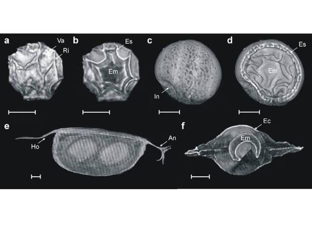
3D reconstructions of resting eggs obtained via X ray scanning. Top left: a cyst of the fairy shrimp Branschipodopsis wolfi, Top right: a cyst of the tadpole shrimp Triops. Bottom: an ephippium with two resting eggs of the water flea Daphnia magna.


